|
Jacob's Journey
Technical notes on Jacob's cancer.
| Here
is a collection of haphazard technical notes that I collected and wrote
during Jacob's cancer treatment. I used it as my way helping make sense
of the information overload that we experienced. I've included it here
on the off-chance it might be useful to someone else. |
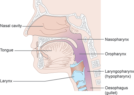 Jacob's tumor (lymphoma) is
located in his posterior nasopharynx.
You can’t see your own nasopharynx directly, but if
you
look inside your mouth in the mirror, it lies above your soft palate
(the soft area at the back of the roof of your mouth) and uvula (the
dangly bit) at the back of the mouth. Jacob's tumor (lymphoma) is
located in his posterior nasopharynx.
You can’t see your own nasopharynx directly, but if
you
look inside your mouth in the mirror, it lies above your soft palate
(the soft area at the back of the roof of your mouth) and uvula (the
dangly bit) at the back of the mouth.
Lymphomas can be found in the nasopharynx. They are cancers of immune
system cells called lymphocytes, cells that are normally found in the
nasopharynx. To understand what lymphoma is, it helps to know
about the body's lymph system.
The lymph system (also known as the lymphatic system) is composed
mainly of lymphoid tissue, lymph vessels, and fluid called lymph (a
clear fluid containing waste products and excess fluid from tissues).
Lymphoid tissue is formed by several types of immune system cells that
work together to help the body fight infections. Lymphoid tissue is
found in many places throughout the body. The adenoids, which
are
a lymphoid tissue, are located in the posterior area of the
nasopharynx and is probably where the cancer started. Most of
the cells found in lymphoid tissue are
lymphocytes, a type of white blood cell. The 2 main types of
lymphocytes are B lymphocytes (B cells) and T lymphocytes (T cells).
Both types can develop into lymphoma cells, but B-cell lymphomas are
much more common than T-cell lymphomas in the United States.
Normal T cells and B cells do different jobs within the immune
system. B cells normally help protect the body against germs
(bacteria or viruses) by making proteins called antibodies. The
antibodies attach to the bacteria or viruses and attract other immune
system cells that surround and digest the antibody-coated germs.
Antibodies also attract certain blood proteins that can kill bacteria.
Lab tests identify B cells and T cells by certain substances on their
surfaces. Some substances are found only on B cells, and others are
found only on T cells. There are also several stages of B-cell and
T-cell development (or maturation) that can be recognized by these lab
tests. This information is helpful because each type of
lymphoma
tends to resemble a particular subtype of normal lymphocytes at a
certain level of development. Determining the type of lymphoma a person
has is the first step in considering treatment options.
There are 2 main types of lymphomas. Hodgkin lymphoma and Non-Hodgkin
lymphoma. These 2 types of lymphoma can usually be
distinguished
from each other by looking at the cancer cells under a microscope. In
some cases, sensitive lab tests may be needed to tell them apart.
Jacob's tumor is a type of cancer known as Burkitt Lymphoma.
This
type makes up about 1% to 2% of all lymphomas. It is named after the
doctor who first described this disease in African children and young
adults. The cells are medium sized and epress the CD20 cell surface
antigen (which is targeted by the chemotherapy drug,
rituximab).
This is a very fast-growing lymphoma. In the African variety, it often
starts as tumors of the jaws or other facial bones. In the more common
types seen in the United States, the lymphoma usually starts in the
abdomen, where it forms a large tumor mass. It can also start in the
ovaries, testes, or other organs, and can spread to the brain and
spinal fluid. Close to 90% of patients are male, and the
average
age is about 30.
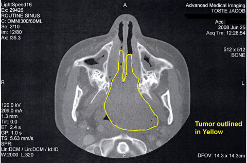
Axial CT slice showing the lymphoma protruding into Jacob's nose.
I was shocked by the size of it (3.7 x 4.1 x 4.9 cm).
After
going through four months of intense chemotherapy, further PET/CT scans
showed that Jacob was in remission. There was a residual polypoid mass
that his physicians suspected was left over scar tissue from the
tumor. On January 8, 2009, five and a half months
after being diagnosed with cancer, Jacob had the residual mass in his
nasopharynx surgically removed. Below are the images captured by
the ENT physician during the procedure:
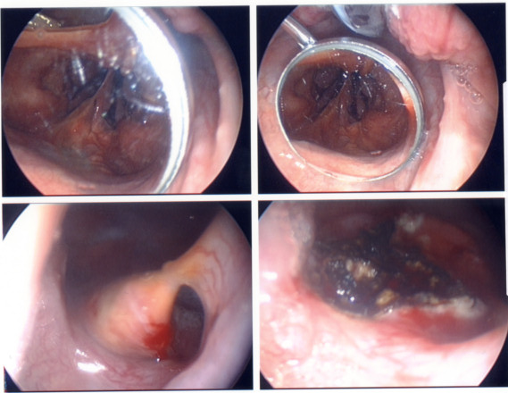
The
top two images show the nasal polyp as seen in a mirror held near the
soft palate. The bottom two images are viewed through the nasal
passage (left image is before excision, right image is after excision
and shows the cauterized area).
|
Jacob's Mediport
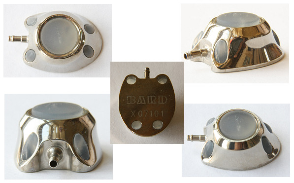
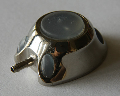
This
is Jacob's actual mediport. We were able to keep it after it was removed. The
device was placed under his skin on his chest. A tube went from the
mediport under his skin, up his chest to his jugular vein, down the
vein and into the right atrium of his heart. It allowed for the best
venous access that would thoroughly mix medication with his blood
thereby reducing possible irritation of his vasculature. Since it was
under his skin it stayed sterile when not in use and reduced the chance
of him picking up an infection.
To learn more about Mediports visit the Wikipedia article.
|
Chemotherapy
The ability of most chemotherapy to kill cancer cells depends on its
ability to halt cell division. Usually, the drugs work by
damaging the RNA or DNA that tells the cell how to copy itself in
division. If the cells are unable to divide, they die. The
faster the cells are dividing, the more likely it is that chemotherapy
will kill the cells, causing the tumor to shrink. They also
induce cell suicide (self-death or apoptosis).
Chemotherapy is most effective at killing cells that are rapidly
dividing. Unfortunately, chemotherapy does not know the
difference between the cancerous cells and the normal cells. The
"normal" cells will grow back and be healthy but in the meantime, side
effects occur. The "normal" cells most commonly affected by
chemotherapy are the blood cells, the cells in the mouth, stomach and
bowel, and the hair follicles; resulting in low blood counts, mouth
sores, nausea, diarrhea, and/or hair loss. Different drugs may
affect different parts of the body.
Methotrexate exerts its chemotherapeutic effect by being able to counteract and
compete with folic acid in cancer cells resulting in folic acid deficiency in
the cells and causing their death. This action also effects normal cells which
can cause significant side effects in the body, such as: low white, red and
platelet blood cell counts, hair loss, mouth sores, difficulty swallowing,
diarrhea, liver, lung, nerve and kidney damage. These complications and side
effects of methotrexate can be either prevented or decreased by using
Leucovorin, which provides a source of folic acid for the body's cells.
Leucovorin is normally started 24 hours after methotrexate is given. This delay
gives the methotrexate a chance to exert its anti cancer effects.
Cytarabine belongs to the category of chemotherapy
called
antimetabolites. Antimetabolites are very similar to normal
substances within the cell (in this case one of the DNA bases).
When the cells incorporate cytarabine into the cellular metabolism,
they are unable to
divide because the drugs acts like a nucleotide and stops DNA
polymerization. Antimetabolites are cell-cycle specific. They
attack cells at very specific phases in the cycle.
|
Hematology 101
Neutrophils (aka polymorphonuclear cells, PMN's, granulocytes,
segmented neutrophils, or segs) fight against infection and
represent a subset of the white blood count. Neutropenia by definition
is an ANC below 1800/mm3 (some sources use a lower value).
-- Absolute neutrophil count (ANC) of 1000-1800: Most patients will be
given chemotherapy in this range. Risk of infection is considered low. (Mild neutropenia)
-- Absolute neutrophil count (ANC) of 500-1000: Carries with it a moderate risk of infection.
-- Absolute neutrophil count (ANC) of less than 500: Severe neutropenia - high risk of infection.
Remember that a reduced WBC is known as leukopenia.
The WBC consists of the following (differential):
Lymphocytes: 20-40%
Neutrophils: 50-60%
Basophils: 0.5-2%
Eosinophils: 1-4%
Monocytes: 2-9% (average: 4%).
The ANC (Absolute Neutrophil Count) refers to the total number of neutrophil granulocytes present in the blood.
ANC = Total WBC x (% "Segs" + % "Bands")
Equivalent to: WBC x ((Segs/100) + (Bands/100))
Normal value: ≥ 1500 cells/mm3.
Mild neutropenia: ≥1000 - <1500/mm3.
Moderate neutropenia: ≥500 - <1000/mm3.
Severe neutropenia: < 500/mm3.
Normal Lab Values
WBC 5.0-14.5 10e3/mcL
RBC 4.0-5.2 10e6/mcL
HGB 11.5-13.5 g/dL
HCT 34-40%
PLT Count 145-400 10e3/mcL
ANC Auto 1500- /mcL
|
|
|


 Jacob's tumor (lymphoma) is
located in his posterior nasopharynx.
You can’t see your own nasopharynx directly, but if
you
look inside your mouth in the mirror, it lies above your soft palate
(the soft area at the back of the roof of your mouth) and uvula (the
dangly bit) at the back of the mouth.
